



Application of desktop scanning electron microscope in the detection of brightness enhancement film of liquid crystal display
Release time:
2023-05-05
Liquid crystal display (Liquid Crystal Display, LCD) is the current social application very much display, in daily life everywhere. LCD is mainly composed of color filter substrate (Color filter,CF), TFT array (TFT Array) substrate and backlight module (Back light unit).
Liquid crystal display (Liquid Crystal Display, LCD) is a display that is widely used in the current society and can be seen everywhere in daily life. LCD is mainly composed of color filter substrate (Color filter,CF), TFT array (TFT Array) substrate and backlight module (Back light unit).
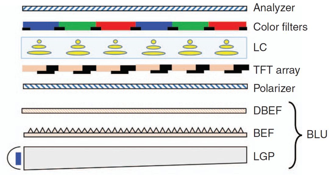
TFT LCD panel structure
(picture comes from network)
In these three components, backlight module is very important to LCD, because backlight quality determines important parameters such as brightness, uniformity of emitted light, color gradation, etc. of LCD screen, and determines the luminous effect of LCD. In the backlight module, there is a very important optical film-brightness enhancement film (Brightness Enhancement Film,BEF). Before the brightness enhancement film was born, the light source utilization rate of the liquid crystal panel was very low. Incremental film, also known as prism sheet (Prism Sheet), originated in the 1990 s and was developed by 3M Company in the United States in the early days. Their technicians applied prism catheter technology to the LCD field. Incremental film is a prism structure formed on the PET surface by using micro-complex technology and thin film technology, and a prism sheet is used to change the optical path of incident light, so that the light emission range is more concentrated and is more effectively used by LCD, thus effectively improving the illumination brightness, uniformity and image contrast, and indirectly improving the power consumption performance of the backlight module. The forming process of

incremental film
(picture comes from the network)
incremental film is mainly divided into two steps: coating film, forming and curing: the PET substrate moves forward, the coating machine evenly covers the surface of the PET substrate with ultraviolet curing adhesive, and then the roller with micro prism structure is formed on the coating layer, at the same time, the ultraviolet lamp is cured to form a microstructure on the surface of the PET substrate and is cured on the surface of the film.
Today we talk about the application of desktop scanning electron microscopy in the characterization of incremental films in LCD backlight modules.
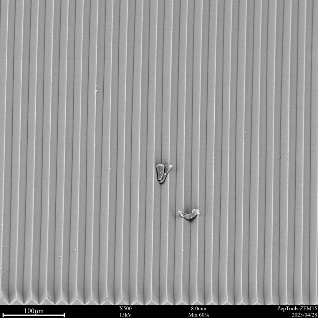
SEM Observation of Prismatic Microstructure on 3M Incremental Film Surface
(BSE & SE Mixed Image BSE 60%,SE accounts for 40%)
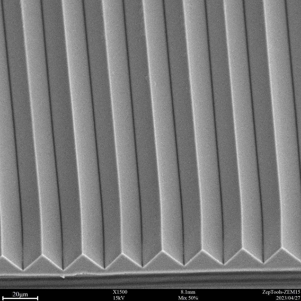
3M incremental film surface morphology structure local enlarged image
(BSE,SE each accounts for 50%)
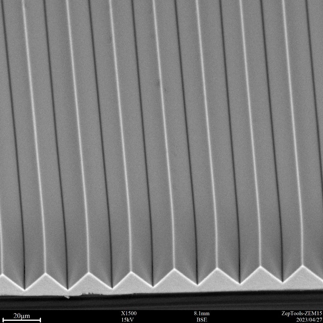
3M incremental film surface microstructure SEM observation, BSE Image
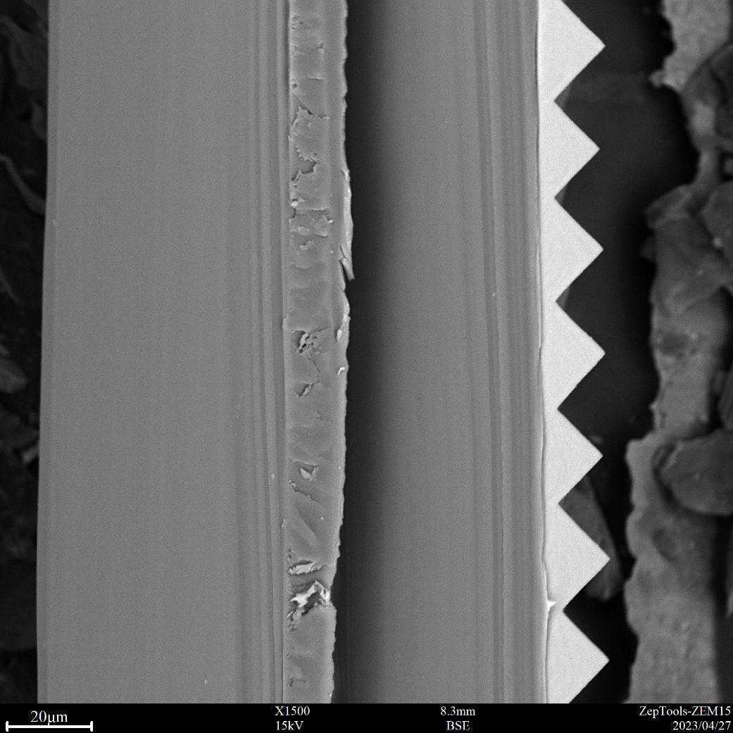
SEM Characterization of Incremental Film Section Morphology
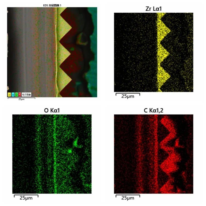
EDS Surface Scan Distribution Image

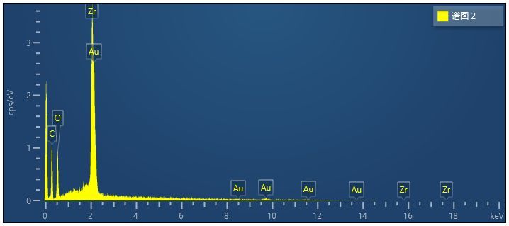
EDS spectrum acquisition of incremental film cross-section sample prismatic structure local area
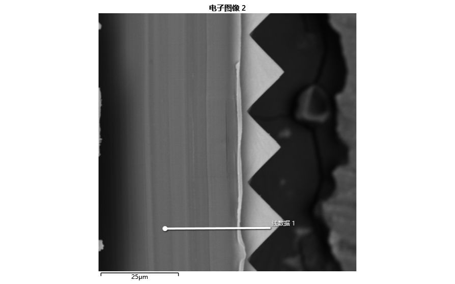
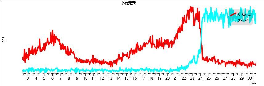
EDS line scan characterization of incremental film cross-section micro-area
We collected EDS spectrum in 3M incremental film cross-section prismatic structure area, it is found that the content of Zr element is higher, the content of C element is higher in the substrate, and the content of C element is not uniformly distributed. The use of EDS line scanning function can be very direct observation of changes in the distribution of chemical elements contained in the material.
In summary: Desktop scanning electron microscope with EDS energy spectrometer is very helpful for microstructure observation and composition analysis of incremental film (prism film).

ZEM15 Desktop Scanning Electron Microscope
Related News
Share

Contact Us
Mobile Phone: 15756003283
Email: support@zeptools.com、micage@zeptools.com
Address: No.15, Electronic Venture Park, Dongshi, Tongling City, Anhui Province, China