



Product classification

ZEM30 Pro Multi-Function SEM
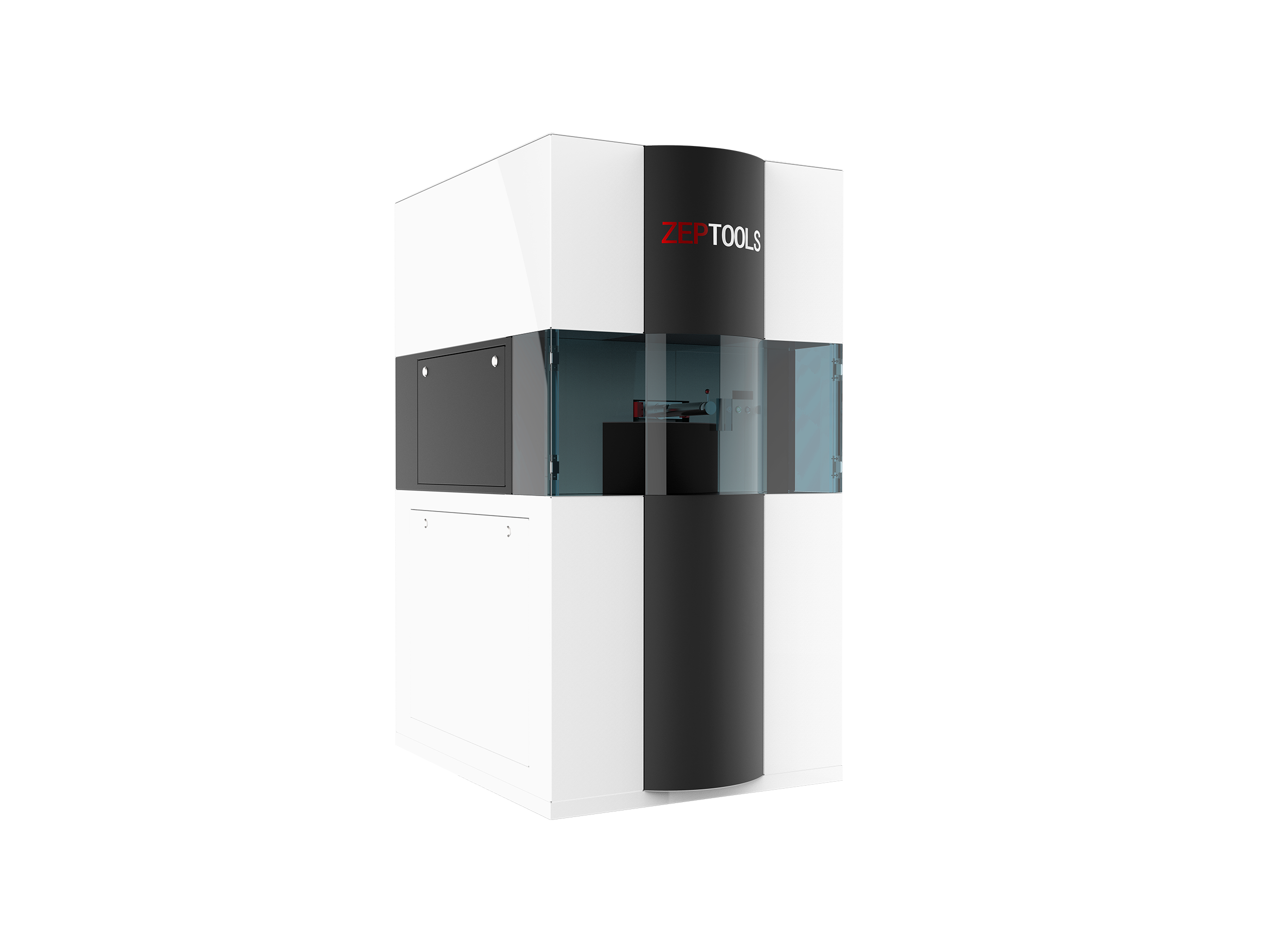
ZEM30 Pro Multi-Function Scanning Electron Microscope
Classification:
Product Description
LaB6 Crystal High-Brightness Filament
ZEM30 PRO
The High-Resolution Choice for General-Purpose Scanning Electron Microscopes
2.5nm Resolution | 500,000x Magnification
Brighter, Clearer
More Powerful Expandability
The ZEM30 Pro is a high-performance general-purpose scanning electron microscope. It breakthroughly adopts a LaB6 (Lanthanum Hexaboride) filament, providing a higher-brightness electron beam source compared to traditional tungsten filaments, thereby significantly improving image quality and pushing resolution to 2.5nm.
More than just observation. The ZEM30 Pro features a modular column design, with a pneumatic structure ensuring the optical path continuously maintains a high vacuum state. Combined with a 5-axis high-precision sample stage and rich interfaces, it can easily accommodate various in-situ stages for tensile, heating, cooling, electrical measurement, and more, making it an all-around platform for micro-nano materials research.
ZEM Series Scanning Electron Microscopes
Full Series Coverage, Choose Your Preferred ZEPTOOLS SEM
| Product Model | ZEM Ultra | ZEM30 Pro | ZEM30 | ZEM20 Pro | ZEM20 | ZEM18 | ZEM10 |
| Resolution | <2.5nm | 2.5nm | 3nm | 3nm | 4nm | <6nm | <10nm |
| Maximum Magnification | 1,000,000x | 500,000x | 500,000x | 500,000x | 360,000x | 360,000x | 250,000x |
| Electron Gun | Schottky Field Emission | LaB6 | Tungsten Filament | LaB6 | Tungsten Filament | Tungsten Filament | Tungsten Filament |
| Accelerating Voltage | 1-15kV Continuously Adjustable | 3-30kV Continuously Adjustable | 3-30kV Continuously Adjustable | 3-20kV Continuously Adjustable (Upgradeable to 30kV) | 3-20kV Continuously Adjustable (Upgradeable to 30kV) | 3-20kV Continuously Adjustable | 5kV/10kV/15kV |
| Detector Type | SE BSE EDS (Optional) | SE BSE EDS (Optional) | SE BSE EDS (Optional) | SE BSE EDS (Optional) | SE BSE EDS (Optional) | SE BSE EDS (Optional) | SE or BSE |
| In-Situ Expansion Capability | Yes | Yes | Yes | Yes | Yes | Yes | —— |
Application Cases
Exceptional Imaging Technology Meets All Your Requirements

More Cases
ZEPTOOLS ZEM Series SEM Helps Research Teams Publish High-Level Journal Achievements


Some Customers
The Trusted Choice of Numerous Universities, Research Institutions, and Enterprises

Start Your High-Resolution Observation Journey
Independently Developed by ZEPTOOLS, Providing a Complete Set of Micro-Nano Analysis Solutions
Recommended Products


Welcome your message consultation

Contact Us
Mobile Phone: 15756003283
Email: support@zeptools.com、micage@zeptools.com
Address: No.15, Electronic Venture Park, Dongshi, Tongling City, Anhui Province, China